


Gonarthrosis of the knee joint is the most common localization of degenerative-dystrophic disease, which is characterized by the gradual destruction of cartilage with subsequent changes in the joint surface, accompanied by pain and reduced mobility.
The disease easily affects women over 40 years old, especially those who are overweight and have varicose veins of the lower extremities.
The knee joint is made up of three compartments:
- tibiofemoral mediators;
- lateral tibiofemoral;
- on the star-thigh.
These compartments can be affected by osteochondrosis (DOA) both individually and in any combination. 75% of all cases of gonarthrosis are destruction of the midbrain cavity (during movement, it is subjected to loads that exceed body weight by 2–3 times).
In young patients, usually only one joint is destroyed - the right or left side (right or left gonarthrosis).
What causes DOA of the knee joint?
Several factors may be involved in the development of concomitant degenerative cartilage changes:
- mechanical overload of the knee joint (some specialties, sports) with microscopic damage of cartilage;
- consequences of trauma, surgical intervention (meniectomy);
- arthritis of the knee (arthritis);
- anatomical inconsistencies of the joint surface (dysplasia);
- static violations (flat feet, curvature of the spine);
- chronic metastatic disease (accumulation of blood in the synovial space);
- metabolic pathologies (gout, hemoglobinopathy, chondrocalcinosis);
- excess body weight;
- violation of the blood supply to the bones;
- osteodystrophy (Paget's disease);
- neurological diseases, loss of sensation in the extremities;
- endocrine disorders (acromegaly, diabetes mellitus, amenorrhea, hyperparathyroidism);
- genetic predisposition (generalized forms of osteoarthritis);
- violation of the synthesis of type II collagen.
But in 40% of cases, the root cause of the disease cannot be identified (primary joint disease).
Pathogenesis of gonarthrosis
early stage
In the early stages of the disease, the metabolic processes of articular cartilage are disturbed. The synthesis and quality of the main structural unit of cartilage tissue, proteoglycans, which are responsible for the stability of the structure of the collagen network, are reduced.
As a result, chondroitin sulfate, keratin, and hyaluronic acid drift out of the mesh, and structurally defective proteoglycans can no longer hold water. It is absorbed into collagen, the fibers swell, leading to a decrease in the cartilage's resistance to stress.
In the synovial cavity, inflammatory substances accumulate, under its action the cartilage is destroyed even faster. Fibrosis of the joint capsule develops. The change in the composition of the synovial fluid makes it difficult to deliver nutrients to the cartilage and interferes with the sliding of the joint surface during movement.
Progression of the disease
In the future, the cartilage gradually becomes thinner, becomes rough, cracks form over its entire thickness. The epigenetic part of bone is subjected to increased loads, leading to the development of osteosclerosis and compensatory proliferation of bone tissues (osteoforms).
This response of the body is intended to increase the joint surface area and redistribute the load. But the presence of osteoclasts increases discomfort, deformity and further limits mobility of the limb.
Microfractures are formed in the thickness of the bone, damaging blood vessels and leading to visceral hypertension. In the final stages of degenerative joint disease, joint surfaces are completely exposed, deformed, and limb movement is severely limited.
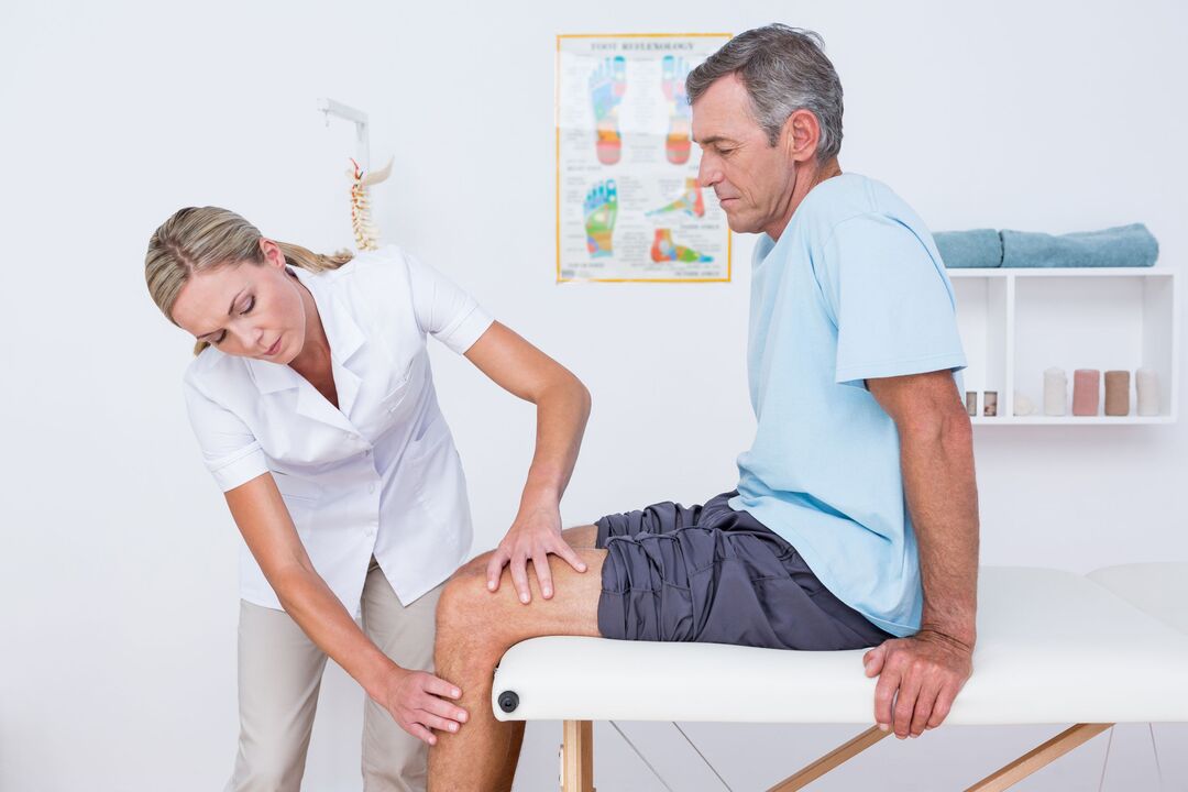
Symptoms of gonarthrosis of the knee joint
Osteoarthritis of the knee is characterized by a chronic, slowly progressive process (months, years). The clinic developed gradually, with no obvious exacerbations. Patients cannot remember exactly when the first symptoms appeared.
Clinical manifestations of gonarthrosis:
- pain. At first, diffuse, short (long standing, walking up stairs), and as osteoarthritis progresses, pain is local (front and inner surface of the knee), their intensity increases;
- Local sensitivity to palpation. Mostly on the medial side of the knee along the edge of the joint space;
- crunch. In stage I it may be inaudible, in stage II-III it accompanies all movements;
- increase in volume, deformity of the knee joint. As a result of weakening of the lateral ligaments, a person develops an O-shaped structure of the limbs (it is clearly visible even in the photo);
- limited mobility. At first there were difficulties with bending the knee, then - with extension.
Causes of pain in DOA:
- mechanical friction of damaged joint surfaces;
- increased intraluminal pressure, venous congestion;
- the accession of bursitis;
- changes in the peristaltic tissues (elongation of the capsule, ligaments, tendons);
- thickening of the periosteum;
- dystrophy of adjacent muscles;
- fibromyalgia;
- compression of nerve endings.
In contrast to coxarthrosis, DOA of the knee may show spontaneously regressing symptoms.
Clinical manifestations of gonarthrosis depend on the stage:
| Characteristics | I stage | Phase II | Phase III |
|---|---|---|---|
| Pain | Short, occurs more often when the knee is extended (long standing, walking up stairs) | Just right, disappears after a night's rest | Pronunciation, disturbing even at night |
| Restrictions on movement | Can't see | There is limited stretching, slight limp. | Persistent, lame-bending contracts |
| crunch | No | Feel it to the touch while moving | cry from afar |
| Distortion | Missing | The axis of the forelimbs is slightly misaligned, losing muscle | Valgus or varus malformation. Unstable joints, atrophy of thigh muscles. |
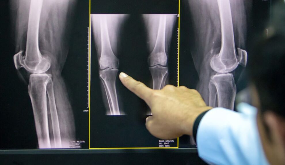
| X-ray images | Mild narrowing of joint space, early sign of subchondral sclerosis | Joint space is narrowed by 50% or more, osteoblasts appear. | Almost complete absence of joint space, significant deformity and hardening of the joint surface, areas of subchondral bone necrosis, osteoporosis |
A common complication of arthritis of the knee joint is secondary reactive bursitis, which is characterized by the following symptoms:
- increasingly painful;
- eye bags;
- effusion into the synovial space;
- increased skin temperature.
Less frequent and more dangerous complications include: joint blockade, femoral necrosis, patellar compression, spontaneous metastasis.
Diagnosis of DOA of the knee joint
The diagnosis of gonarthrosis is based on the characteristic complaints of the patient, changes detected during examination, and the results of additional tests.
To confirm osteoarthritis, it is prescribed:
- X-ray of the knee joint in two views (anterior and posterior): the most accessible way to confirm the diagnosis in the advanced stages of the disease;
- Ultrasound: determine the presence of effusion in the joint, measure the cartilage thickness;
- synovial fluid analysis;
- diagnostic arthroscopy (visual assessment of cartilage) with biopsy;
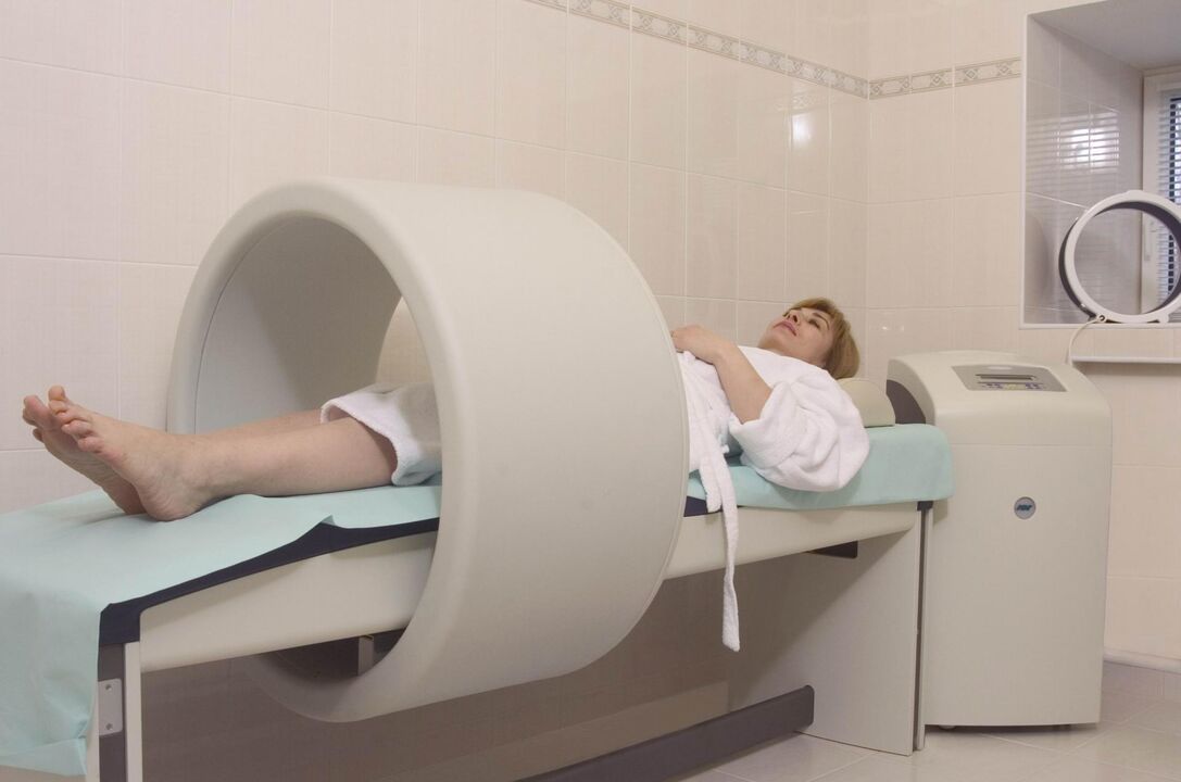
- Computer and magnetic resonance imaging (CT, MRI): the best method to diagnose DOA in the early stages.
If the doctor is in doubt about the diagnosis, it may be prescribed:
- scintigraphy: joint scan after the introduction of radioisotopes;
- temperature: study the intensity of infrared radiation (its strength is proportional to the strength of inflammation).
Treatment of gonarthrosis of the knee joint
The treatment regimen for knee osteoarthritis combines many methods: non-drug methods, drugs and corrective surgery. The proportions of each method were determined individually for each patient.
Non-drug treatment
In the latest ESCEO (European Society for Clinical Aspects of Osteoporosis and Osteoarthritis) guidelines on the treatment of knee osteoarthritis, the experts place particular emphasis on patient education andlifestyle change.
The patient needs:
- explain what the nature of the disease is, established for long-term treatment;
- teach how to use assistive devices (canes, orthotics);
- prescribe a diet (for patients with a body mass index above 30);
- do exercises to strengthen the thigh muscles and unload the knee joint;
- Explain the importance of increasing physical activity.
In the early stages of knee effusion, treatment with physiotherapy gives good results:
- Massage;
- acupuncture therapy;
- UHF therapy;
- electrophoresis;
- hydrogen sulfide bath;
- paraffin application;
- Acupuncture.
Pharmacotherapeutic gonarthrosis
The use of drugs in DOA is aimed at reducing pain, reducing inflammation, and slowing the rate of cartilage destruction.
Symptomatic treatment:
- analgesic;
- non-steroidal anti-inflammatory drugs (NSAIDs) of the COX-2 inhibitor class in the form of tablets or suppositories;
- non-narcotic pain relievers (with drug-resistant pain syndrome).
Chondroprotectors:
- Chondroitin sulfate;
- Glucosamine sulfate.
These drugs can be taken in the form of capsules in courses several times a year, intramuscularly or directly into the synovial cavity.
Topical therapy includes proximal and intra-articular injections of glucocorticosteroids, hyaluronic acid preparations.
In stages I-II of DOA, an important place in complex therapy is the use of NSAID-based anti-inflammatory ointments, gels and creams. They help reduce the patient's need for oral NSAIDs, thereby reducing the risk of gastrointestinal damage.
Folk methods of treatment
The use of decoction, decoction, extract, topical application of medicinal plants should be considered as an adjunct to the treatment of DOA, folk remedies cannot replace the therapy prescribed by a doctor. .
Plants used in osteoarthritis: dandelion, ginger, Jerusalem artichoke, burdock, garlic, sea buckthorn.
Surgery
Surgical intervention may be required at all stages of gonadal disease without sufficient effect of medical measures. The most common is the laparoscopic procedure, in the most severe cases organ replacement is indicated.
Types of endoscopic interventions:
- joint adjustment and restoration: extraction of inflammatory substances from the synovial cavity, fragments of cartilage;
- plasma or laser ablation: removal of mechanical obstructions in the synovial cavity;
- chondroplasty.
Peristaltic osteotomy is indicated for patients whose initial presentation is axial deformity (no more than 15–20%).
The purpose of the operation is to restore the normal configuration of the joint, evenly distribute the load on the joint surface and eliminate damaged areas. This procedure allows you to delay the arthroplasty.
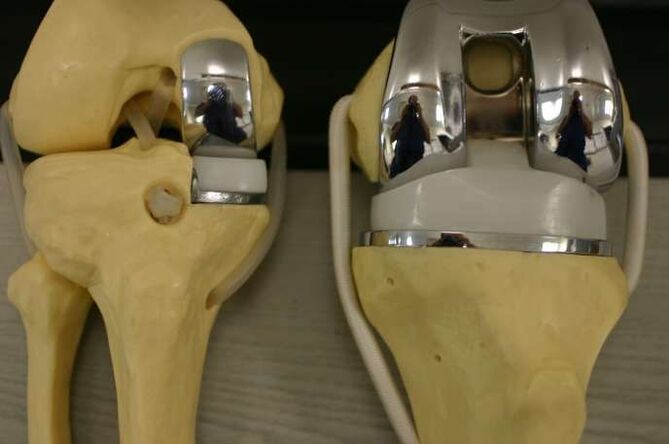
Indications to replace the affected area (or the entire joint) with an artificial one:
- DOA grade II-III;
- severe axial deformity of the limb;
- aseptic necrosis of the subchondral bone layer;
- persistent pain syndrome.
Contraindications for knee orthopedics:
- total damage to joints;
- unstable ligament apparatus;
- DOA as a result of arthritis;
- persistent muscle spasticity, severe muscle weakness.
In this case, the patient underwent knee arthroplasty - comparing the knee joint in physiological position with the removal of the joint surfaces. This relieves pain but shortens the leg, causing secondary damage to the lateral knee, hip, and spine.
Prevent
Prevention of early cartilage degeneration should begin at an early age.
Preventive measures:
- prevention of curvature of the spine;
- flat foot correction (shoe with arch support);
- regular physical education (limited playing heavy sports);
- exclude fixed postures during work.